Choledochal Cyst
Published: 18 Jun 2025
ICD9: 751.69 ICD10: Q44.4 ICD11: LB20.20
A choledochal cyst is a rare, congenital condition involving an abnormal cystic dilation (ballooning) of the biliary tree, specifically the bile ducts.
Here's a breakdown of what that means:
![]() Congenital: This means it's present at birth, although it may not be diagnosed until later in life.
Congenital: This means it's present at birth, although it may not be diagnosed until later in life.![]() Biliary Tree: This is the network of ducts that carry bile from the liver and gallbladder to the small intestine. Bile is a fluid that helps digest fats.
Biliary Tree: This is the network of ducts that carry bile from the liver and gallbladder to the small intestine. Bile is a fluid that helps digest fats.![]() Choledochal: Refers to the common bile duct, which is a major duct in the biliary tree.
Choledochal: Refers to the common bile duct, which is a major duct in the biliary tree.![]() Cystic Dilation: This means an abnormal, pouch-like widening or expansion of the bile duct. It's like a balloon inflating in the bile duct system.
Cystic Dilation: This means an abnormal, pouch-like widening or expansion of the bile duct. It's like a balloon inflating in the bile duct system.
In simpler terms: Imagine the bile ducts as a series of pipes carrying fluid. In choledochal cysts, one of those pipes (usually the common bile duct) has a widened, ballooned-out section, forming a cyst.
Types of Choledochal Cysts:
The Todani classification system is commonly used to categorize choledochal cysts into five types:![]() Type I: The most common type. It involves a fusiform (spindle-shaped) dilation of the common bile duct.
Type I: The most common type. It involves a fusiform (spindle-shaped) dilation of the common bile duct.![]() Type II: A diverticulum (outpouching) arising from the common bile duct.
Type II: A diverticulum (outpouching) arising from the common bile duct.![]() Type III (Choledochocele): Dilation of the distal (end) part of the common bile duct within the wall of the duodenum (the first part of the small intestine).
Type III (Choledochocele): Dilation of the distal (end) part of the common bile duct within the wall of the duodenum (the first part of the small intestine).![]() Type IVa: Multiple dilations involving both the intrahepatic (inside the liver) and extrahepatic (outside the liver) bile ducts.
Type IVa: Multiple dilations involving both the intrahepatic (inside the liver) and extrahepatic (outside the liver) bile ducts.![]() Type IVb: Multiple dilations involving only the extrahepatic bile ducts.
Type IVb: Multiple dilations involving only the extrahepatic bile ducts.![]() Type V (Caroli's disease): Multiple dilations of the intrahepatic bile ducts.
Type V (Caroli's disease): Multiple dilations of the intrahepatic bile ducts.
Causes:
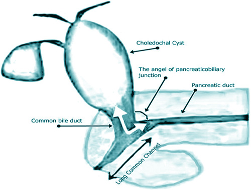
The exact cause isn't fully understood. However, the most accepted theory involves an abnormal junction between the common bile duct and the pancreatic duct (the duct that carries pancreatic enzymes). This abnormal junction allows pancreatic enzymes to reflux into the bile duct, causing inflammation and weakening of the bile duct walls, eventually leading to cyst formation.
Symptoms:
Symptoms can vary depending on the type and size of the cyst, and the age of the patient. They may include:![]() Abdominal pain: Often in the upper right quadrant.
Abdominal pain: Often in the upper right quadrant.![]() Jaundice: Yellowing of the skin and eyes.
Jaundice: Yellowing of the skin and eyes.![]() Palpable abdominal mass: A lump that can be felt in the abdomen (more common in infants).
Palpable abdominal mass: A lump that can be felt in the abdomen (more common in infants).![]() Cholangitis: Inflammation of the bile ducts, leading to fever, chills, and abdominal pain.
Cholangitis: Inflammation of the bile ducts, leading to fever, chills, and abdominal pain.![]() Pancreatitis: Inflammation of the pancreas.
Pancreatitis: Inflammation of the pancreas.
Diagnosis:
Diagnosis typically involves:![]() Ultrasound: Often the first imaging test used.
Ultrasound: Often the first imaging test used.![]() CT scan (Computed Tomography): Provides more detailed images.
CT scan (Computed Tomography): Provides more detailed images.![]() MRI (Magnetic Resonance Imaging) with MRCP (MR Cholangiopancreatography): Provides detailed images of the biliary and pancreatic ducts.
MRI (Magnetic Resonance Imaging) with MRCP (MR Cholangiopancreatography): Provides detailed images of the biliary and pancreatic ducts.![]() ERCP (Endoscopic Retrograde Cholangiopancreatography): An invasive procedure that allows direct visualization of the bile ducts and pancreatic duct (used less frequently for diagnosis but can be used for treatment).
ERCP (Endoscopic Retrograde Cholangiopancreatography): An invasive procedure that allows direct visualization of the bile ducts and pancreatic duct (used less frequently for diagnosis but can be used for treatment).
Treatment:
The primary treatment is surgical removal of the cyst. The dilated portion of the bile duct is resected, and the remaining bile duct is reconnected to the small intestine (usually a Roux-en-Y hepaticojejunostomy). Surgery is important to prevent complications, especially the increased risk of bile duct cancer later in life.
Complications if left untreated:![]() Cholangitis (infection of the bile ducts)
Cholangitis (infection of the bile ducts)![]() Biliary cirrhosis (scarring of the liver)
Biliary cirrhosis (scarring of the liver)![]() Pancreatitis (inflammation of the pancreas)
Pancreatitis (inflammation of the pancreas)![]() Bile duct cancer (cholangiocarcinoma)
Bile duct cancer (cholangiocarcinoma)
In Summary:
A choledochal cyst is a rare birth defect where a portion of the bile duct abnormally widens. It's important to diagnose and treat it surgically to prevent serious complications, including cancer.