Gastrointestinal stromal tumors (GIST)
Published: 18 Jun 2025
ICD9: 171.5 ICD10: C49.A0 ICD11: XH9HQ1
Gastrointestinal stromal tumors (GISTs) are a type of sarcoma, a rare cancer that originates in the soft tissues of the body.
Specifically, GISTs develop in the specialized nerve cells found in the wall of the digestive tract, called the interstitial cells of Cajal (ICCs). These ICCs help control the movement of food and liquid through the digestive system.
Here's a breakdown of important aspects of GISTs:
![]() Origin: Arise from ICCs in the wall of the gastrointestinal (GI) tract.
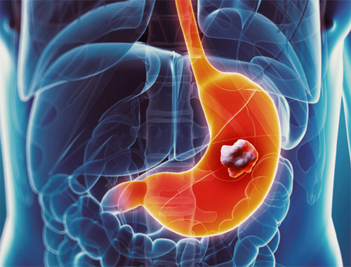
Origin: Arise from ICCs in the wall of the gastrointestinal (GI) tract.![]() Location: Can occur anywhere along the GI tract, but are most common in the stomach (60-70%) and small intestine (20-30%). Less common locations include the esophagus, colon, rectum, and rarely outside the GI tract.
Location: Can occur anywhere along the GI tract, but are most common in the stomach (60-70%) and small intestine (20-30%). Less common locations include the esophagus, colon, rectum, and rarely outside the GI tract.![]() Genetic Mutation: Most GISTs are caused by a mutation in the *KIT* gene (about 75-80% of cases) or the *PDGFRA* gene (about 5-10% of cases). These mutations cause the cells to grow and divide uncontrollably.
Genetic Mutation: Most GISTs are caused by a mutation in the *KIT* gene (about 75-80% of cases) or the *PDGFRA* gene (about 5-10% of cases). These mutations cause the cells to grow and divide uncontrollably.![]() Behavior: GISTs can be benign (non-cancerous) or malignant (cancerous). Even those that appear benign can sometimes recur or spread (metastasize) after removal. Therefore, all GISTs are considered to have some degree of malignant potential. The size of the tumor, its growth rate, and the mitotic rate (how quickly the cells are dividing) help determine the risk of recurrence or metastasis.
Behavior: GISTs can be benign (non-cancerous) or malignant (cancerous). Even those that appear benign can sometimes recur or spread (metastasize) after removal. Therefore, all GISTs are considered to have some degree of malignant potential. The size of the tumor, its growth rate, and the mitotic rate (how quickly the cells are dividing) help determine the risk of recurrence or metastasis.![]() Symptoms: Symptoms vary depending on the size and location of the tumor. Small GISTs may not cause any symptoms. Larger tumors can cause:
Symptoms: Symptoms vary depending on the size and location of the tumor. Small GISTs may not cause any symptoms. Larger tumors can cause:![]()

![]() Abdominal pain or discomfort
Abdominal pain or discomfort![]()
![]() Nausea and vomiting
Nausea and vomiting![]()
![]() Feeling full after eating only a small amount of food
Feeling full after eating only a small amount of food![]()
![]() Loss of appetite
Loss of appetite![]()
![]() Fatigue
Fatigue![]()
![]() Anemia (due to bleeding in the GI tract)
Anemia (due to bleeding in the GI tract)![]()
![]() Blood in the stool or vomit
Blood in the stool or vomit![]() Diagnosis: Diagnosis typically involves:
Diagnosis: Diagnosis typically involves:![]()
![]() Endoscopy: A procedure where a thin, flexible tube with a camera is inserted into the GI tract to visualize the lining.
Endoscopy: A procedure where a thin, flexible tube with a camera is inserted into the GI tract to visualize the lining.![]()
![]() Imaging scans: CT scans, MRI scans, and PET scans can help identify the tumor and assess its size and spread.
Imaging scans: CT scans, MRI scans, and PET scans can help identify the tumor and assess its size and spread.![]()
![]() Biopsy: A tissue sample is taken from the tumor and examined under a microscope to confirm the diagnosis and determine the type of cells involved. Immunohistochemistry (IHC) is a special test that helps identify specific proteins on the cells, such as KIT (CD117) or DOG1, which are commonly found in GISTs.
Biopsy: A tissue sample is taken from the tumor and examined under a microscope to confirm the diagnosis and determine the type of cells involved. Immunohistochemistry (IHC) is a special test that helps identify specific proteins on the cells, such as KIT (CD117) or DOG1, which are commonly found in GISTs.![]() Treatment: Treatment depends on the size, location, and risk of recurrence or metastasis. Options include:
Treatment: Treatment depends on the size, location, and risk of recurrence or metastasis. Options include:![]()
![]() Surgery: The primary treatment for localized GISTs.
Surgery: The primary treatment for localized GISTs.![]()
![]() Targeted therapy: Drugs that specifically target the *KIT* or *PDGFRA* proteins, such as imatinib (Gleevec), sunitinib (Sutent), and regorafenib (Stivarga). These drugs are often used after surgery to prevent recurrence or to treat metastatic disease. Avapritinib (Ayvakit) and ripretinib (Qinlock) are used for specific mutations.
Targeted therapy: Drugs that specifically target the *KIT* or *PDGFRA* proteins, such as imatinib (Gleevec), sunitinib (Sutent), and regorafenib (Stivarga). These drugs are often used after surgery to prevent recurrence or to treat metastatic disease. Avapritinib (Ayvakit) and ripretinib (Qinlock) are used for specific mutations.![]()
![]() Radiation therapy: Less commonly used than surgery or targeted therapy, but may be used in certain situations.
Radiation therapy: Less commonly used than surgery or targeted therapy, but may be used in certain situations.![]() Prognosis: The prognosis for GISTs varies widely depending on the size, location, and risk of recurrence or metastasis. Localized GISTs that are completely removed surgically have a good prognosis. Metastatic GISTs are more challenging to treat, but targeted therapy has significantly improved survival rates.
Prognosis: The prognosis for GISTs varies widely depending on the size, location, and risk of recurrence or metastasis. Localized GISTs that are completely removed surgically have a good prognosis. Metastatic GISTs are more challenging to treat, but targeted therapy has significantly improved survival rates.
In summary, GISTs are tumors that arise from specialized cells in the GI tract wall. They are often caused by genetic mutations and can be treated with surgery and/or targeted therapy. Early diagnosis and treatment are important for improving outcomes.