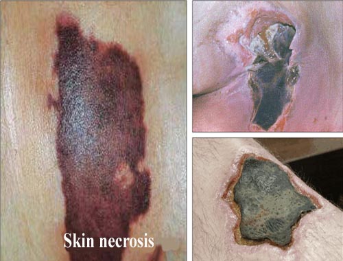
Necrosis

Published: 18 Jun 2025
ICD9: 733.40 ICD10: R02.0 ICD11: MC85
Necrosis is the death of cells or tissues within a living organism.
It's a type of cell death that's often caused by external factors such as infection, toxins, trauma, or ischemia (lack of blood supply).
Here's a breakdown of what's important to understand about necrosis:
![]() It's Premature Death: Necrosis is different from apoptosis (programmed cell death), which is a natural and controlled process for removing unwanted or damaged cells. Necrosis is more like an accidental death, often occurring in response to harmful conditions.
It's Premature Death: Necrosis is different from apoptosis (programmed cell death), which is a natural and controlled process for removing unwanted or damaged cells. Necrosis is more like an accidental death, often occurring in response to harmful conditions.
![]() Causes: Many things can trigger necrosis, including:
Causes: Many things can trigger necrosis, including:![]()

![]() Ischemia: Lack of oxygen due to blocked blood flow (e.g., in a heart attack or stroke)
Ischemia: Lack of oxygen due to blocked blood flow (e.g., in a heart attack or stroke)![]()

![]() Infection: Bacterial, viral, or fungal infections can directly damage cells or release toxins that cause necrosis.
Infection: Bacterial, viral, or fungal infections can directly damage cells or release toxins that cause necrosis.![]()

![]() Toxins: Exposure to chemicals, poisons, or drugs can be toxic to cells and lead to necrosis.
Toxins: Exposure to chemicals, poisons, or drugs can be toxic to cells and lead to necrosis.![]()

![]() Trauma: Physical injury, burns, or frostbite can directly damage tissues.
Trauma: Physical injury, burns, or frostbite can directly damage tissues.![]()

![]() Radiation: Exposure to high doses of radiation can kill cells.
Radiation: Exposure to high doses of radiation can kill cells.![]()

![]() Immunological Injury: In some autoimmune diseases, the immune system mistakenly attacks and destroys healthy cells, leading to necrosis.
Immunological Injury: In some autoimmune diseases, the immune system mistakenly attacks and destroys healthy cells, leading to necrosis.
![]() Cellular Changes: When a cell undergoes necrosis, it experiences several changes:
Cellular Changes: When a cell undergoes necrosis, it experiences several changes:![]()

![]() Cell Swelling: The cell enlarges due to an influx of water and ions.
Cell Swelling: The cell enlarges due to an influx of water and ions.![]()

![]() Membrane Rupture: The cell membrane breaks down, releasing its contents into the surrounding tissue.
Membrane Rupture: The cell membrane breaks down, releasing its contents into the surrounding tissue.![]()

![]() Inflammation: The release of cellular contents triggers an inflammatory response, attracting immune cells to the area.
Inflammation: The release of cellular contents triggers an inflammatory response, attracting immune cells to the area.![]()

![]() DNA Degradation: The cell's DNA breaks down.
DNA Degradation: The cell's DNA breaks down.
![]() Types of Necrosis: There are different types of necrosis, characterized by their appearance and the underlying cause:
Types of Necrosis: There are different types of necrosis, characterized by their appearance and the underlying cause:![]()

![]() Coagulative Necrosis: This is the most common type and is often seen in ischemia. The tissue becomes firm and opaque.
Coagulative Necrosis: This is the most common type and is often seen in ischemia. The tissue becomes firm and opaque.![]()

![]() Liquefactive Necrosis: The tissue becomes liquid due to enzymatic digestion. This is common in brain infarcts and bacterial infections.
Liquefactive Necrosis: The tissue becomes liquid due to enzymatic digestion. This is common in brain infarcts and bacterial infections.![]()

![]() Caseous Necrosis: This type is characteristic of tuberculosis. The tissue has a cheese-like appearance.
Caseous Necrosis: This type is characteristic of tuberculosis. The tissue has a cheese-like appearance.![]()

![]() Fat Necrosis: This occurs when fat tissue is damaged, often due to trauma or pancreatitis.
Fat Necrosis: This occurs when fat tissue is damaged, often due to trauma or pancreatitis.![]()

![]() Fibrinoid Necrosis: This is seen in blood vessel walls and is characterized by the deposition of fibrin-like material.
Fibrinoid Necrosis: This is seen in blood vessel walls and is characterized by the deposition of fibrin-like material.![]()

![]() Gangrenous Necrosis: A clinical term, usually used to describe necrosis with superimposed bacterial infection, often in a limb. Dry gangrene is like coagulative necrosis, while wet gangrene is like liquefactive necrosis.
Gangrenous Necrosis: A clinical term, usually used to describe necrosis with superimposed bacterial infection, often in a limb. Dry gangrene is like coagulative necrosis, while wet gangrene is like liquefactive necrosis.
![]() Consequences: Necrosis can have serious consequences for the body, including:
Consequences: Necrosis can have serious consequences for the body, including:![]()

![]() Inflammation: The inflammatory response can damage surrounding healthy tissues.
Inflammation: The inflammatory response can damage surrounding healthy tissues.![]()

![]() Scarring: Dead tissue may be replaced by scar tissue, which can impair organ function.
Scarring: Dead tissue may be replaced by scar tissue, which can impair organ function.![]()

![]() Infection: Necrotic tissue can become a breeding ground for bacteria, leading to infection.
Infection: Necrotic tissue can become a breeding ground for bacteria, leading to infection.![]()

![]() Systemic Effects: In severe cases, necrosis can trigger a systemic inflammatory response (sepsis) and even death.
Systemic Effects: In severe cases, necrosis can trigger a systemic inflammatory response (sepsis) and even death.
![]() Diagnosis: Necrosis can be diagnosed through various methods, including:
Diagnosis: Necrosis can be diagnosed through various methods, including:![]()

![]() Physical Examination: Observing the affected area for signs of tissue damage.
Physical Examination: Observing the affected area for signs of tissue damage.![]()

![]() Imaging Studies: X-rays, CT scans, MRI scans, and ultrasound can help visualize necrotic tissue.
Imaging Studies: X-rays, CT scans, MRI scans, and ultrasound can help visualize necrotic tissue.![]()

![]() Biopsy: A tissue sample can be examined under a microscope to confirm the presence of necrosis and determine its type.
Biopsy: A tissue sample can be examined under a microscope to confirm the presence of necrosis and determine its type.![]()

![]() Blood Tests: Certain blood markers can indicate tissue damage.
Blood Tests: Certain blood markers can indicate tissue damage.
![]() Treatment: Treatment for necrosis depends on the underlying cause and the extent of the damage. Options may include:
Treatment: Treatment for necrosis depends on the underlying cause and the extent of the damage. Options may include:![]()

![]() Treating the Underlying Cause: Addressing the infection, restoring blood flow, or removing toxins.
Treating the Underlying Cause: Addressing the infection, restoring blood flow, or removing toxins.![]()

![]() Debridement: Removing dead tissue to prevent infection and promote healing.
Debridement: Removing dead tissue to prevent infection and promote healing.![]()

![]() Antibiotics: Treating bacterial infections.
Antibiotics: Treating bacterial infections.![]()

![]() Surgery: In severe cases, amputation may be necessary to remove necrotic tissue.
Surgery: In severe cases, amputation may be necessary to remove necrotic tissue.![]()

![]() Hyperbaric Oxygen Therapy: Increasing oxygen levels in the blood to promote healing.
Hyperbaric Oxygen Therapy: Increasing oxygen levels in the blood to promote healing.
In summary, necrosis is a pathological process involving the death of cells and tissues within a living organism due to external factors. It can have serious consequences and requires prompt diagnosis and treatment.