Pneumothorax
Published: 18 Jun 2025
ICD9: 512.89 ICD10: J93.9 ICD11: CB21
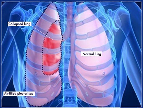
A pneumothorax, often referred to as a collapsed lung, is a condition where air leaks into the space between your lung and chest wall.
This space, called the pleural space, is normally filled with a thin layer of fluid that allows the lung to expand and contract smoothly with each breath. When air enters this space, it puts pressure on the lung, causing it to collapse, either partially or completely.
Here's a breakdown of the key aspects:
![]() What happens: Air enters the pleural space (the space between the lung and chest wall).
What happens: Air enters the pleural space (the space between the lung and chest wall).![]() Why it matters: This air pressure prevents the lung from fully expanding.
Why it matters: This air pressure prevents the lung from fully expanding.![]() Result: The lung collapses.
Result: The lung collapses.
Types of Pneumothorax:![]() Spontaneous Pneumothorax: This occurs without any obvious injury. It is further divided into:
Spontaneous Pneumothorax: This occurs without any obvious injury. It is further divided into:![]()

![]() *Primary Spontaneous Pneumothorax:* Occurs in people without underlying lung disease, often tall, thin young men. Blisters (blebs) on the top of the lung can rupture and leak air.
*Primary Spontaneous Pneumothorax:* Occurs in people without underlying lung disease, often tall, thin young men. Blisters (blebs) on the top of the lung can rupture and leak air.![]()
![]() *Secondary Spontaneous Pneumothorax:* Occurs in people with underlying lung disease, such as COPD, asthma, cystic fibrosis, or lung cancer.
*Secondary Spontaneous Pneumothorax:* Occurs in people with underlying lung disease, such as COPD, asthma, cystic fibrosis, or lung cancer.![]() Traumatic Pneumothorax: This is caused by an injury to the chest, such as:
Traumatic Pneumothorax: This is caused by an injury to the chest, such as:![]()
![]() A penetrating wound (e.g., gunshot or stab wound).
A penetrating wound (e.g., gunshot or stab wound).![]()
![]() A blunt force trauma (e.g., car accident or fall).
A blunt force trauma (e.g., car accident or fall).![]()
![]() Medical procedures, like lung biopsy or central line insertion.
Medical procedures, like lung biopsy or central line insertion.![]() Tension Pneumothorax: This is a life-threatening condition where air enters the pleural space but cannot escape. The pressure in the chest rapidly increases, compressing the lung and heart, leading to decreased blood pressure and oxygenation. It is a medical emergency.
Tension Pneumothorax: This is a life-threatening condition where air enters the pleural space but cannot escape. The pressure in the chest rapidly increases, compressing the lung and heart, leading to decreased blood pressure and oxygenation. It is a medical emergency.
Causes:![]() Lung disease: COPD, asthma, cystic fibrosis, pneumonia, lung cancer.
Lung disease: COPD, asthma, cystic fibrosis, pneumonia, lung cancer.![]() Injury: Trauma to the chest.
Injury: Trauma to the chest.![]() Ruptured air blisters (blebs): Especially in tall, thin young adults.
Ruptured air blisters (blebs): Especially in tall, thin young adults.![]() Medical procedures: Lung biopsy, central line insertion, mechanical ventilation.
Medical procedures: Lung biopsy, central line insertion, mechanical ventilation.
Symptoms:![]() Sudden chest pain
Sudden chest pain![]() Shortness of breath
Shortness of breath![]() Cough
Cough![]() Fatigue
Fatigue![]() Rapid heart rate
Rapid heart rate![]() Bluish discoloration of the skin (cyanosis) - in severe cases
Bluish discoloration of the skin (cyanosis) - in severe cases
Diagnosis:![]() Physical exam
Physical exam![]() Chest X-ray
Chest X-ray![]() CT scan
CT scan
Treatment:
The treatment depends on the size of the pneumothorax and the severity of symptoms:![]() Observation: Small pneumothoraxes may resolve on their own.
Observation: Small pneumothoraxes may resolve on their own.![]() Needle aspiration: Using a needle to remove air from the pleural space.
Needle aspiration: Using a needle to remove air from the pleural space.![]() Chest tube insertion: Inserting a tube into the chest to drain air and allow the lung to re-expand.
Chest tube insertion: Inserting a tube into the chest to drain air and allow the lung to re-expand.![]() Surgery: In some cases, surgery may be needed to repair air leaks or remove damaged lung tissue. Pleurodesis, a procedure to adhere the lung to the chest wall, may also be performed to prevent recurrence.
Surgery: In some cases, surgery may be needed to repair air leaks or remove damaged lung tissue. Pleurodesis, a procedure to adhere the lung to the chest wall, may also be performed to prevent recurrence.
In summary, a pneumothorax is a potentially serious condition that requires prompt medical attention. Understanding the different types, causes, symptoms, and treatment options is crucial for effective management.