Renal cell carcinoma
Published: 18 Jun 2025
ICD9: 189.0 ICD10: C64.9 ICD11: XH05V6
Renal cell carcinoma (RCC) is a type of kidney cancer that originates in the lining of the proximal convoluted tubule, the part of the kidney that filters the blood and removes waste products.
It's the most common type of kidney cancer in adults, accounting for about 85% of kidney malignancies.
Here's a breakdown of important aspects of renal cell carcinoma:
Key Features:
![]() Origin: Arises from the cells lining the kidney tubules (primarily the proximal convoluted tubule).
Origin: Arises from the cells lining the kidney tubules (primarily the proximal convoluted tubule).![]() Prevalence: Most common type of kidney cancer in adults.
Prevalence: Most common type of kidney cancer in adults.![]() Typically occurs: Usually develops as a single tumor in one kidney, but can occur in both kidneys or as multiple tumors.
Typically occurs: Usually develops as a single tumor in one kidney, but can occur in both kidneys or as multiple tumors.
Causes and Risk Factors:
While the exact cause of RCC is often unknown, several factors increase the risk:![]() Smoking: A major risk factor.
Smoking: A major risk factor.![]() Obesity: Increased risk due to hormonal and metabolic changes.
Obesity: Increased risk due to hormonal and metabolic changes.![]() High blood pressure (Hypertension): Long-term hypertension is linked to increased risk.
High blood pressure (Hypertension): Long-term hypertension is linked to increased risk.![]() Family history: Having a close relative with kidney cancer increases the risk.
Family history: Having a close relative with kidney cancer increases the risk.![]() Certain genetic conditions: Von Hippel-Lindau (VHL) disease, Birt-Hogg-Dube syndrome, hereditary papillary renal cell carcinoma, and tuberous sclerosis complex are associated with higher RCC risk.
Certain genetic conditions: Von Hippel-Lindau (VHL) disease, Birt-Hogg-Dube syndrome, hereditary papillary renal cell carcinoma, and tuberous sclerosis complex are associated with higher RCC risk.![]() Chronic kidney disease and Dialysis: Long-term dialysis treatment is associated with increased risk.
Chronic kidney disease and Dialysis: Long-term dialysis treatment is associated with increased risk.![]() Exposure to certain chemicals: Such as trichloroethylene (TCE) and cadmium.
Exposure to certain chemicals: Such as trichloroethylene (TCE) and cadmium.![]() Age: Risk increases with age, typically diagnosed between ages 60 and 70.
Age: Risk increases with age, typically diagnosed between ages 60 and 70.![]() Race/Ethnicity: African Americans have a slightly higher incidence of RCC than Caucasians.
Race/Ethnicity: African Americans have a slightly higher incidence of RCC than Caucasians.![]() Male gender: Men are more likely to develop RCC than women.
Male gender: Men are more likely to develop RCC than women.
Types of Renal Cell Carcinoma:
There are several subtypes of RCC, classified by their appearance under a microscope. The most common types include:![]() Clear Cell RCC: The most common subtype (70-80% of cases). Cells appear clear under the microscope due to high lipid content. Often associated with VHL gene mutations.
Clear Cell RCC: The most common subtype (70-80% of cases). Cells appear clear under the microscope due to high lipid content. Often associated with VHL gene mutations.![]() Papillary RCC: The second most common subtype (10-15% of cases). Characterized by finger-like projections (papillae). Often associated with MET gene mutations.
Papillary RCC: The second most common subtype (10-15% of cases). Characterized by finger-like projections (papillae). Often associated with MET gene mutations.![]() Chromophobe RCC: (About 5% of cases). Has a better prognosis than clear cell and papillary RCC.
Chromophobe RCC: (About 5% of cases). Has a better prognosis than clear cell and papillary RCC.![]() Collecting Duct RCC: Rare and aggressive.
Collecting Duct RCC: Rare and aggressive.![]() Unclassified RCC: Does not fit into the other categories.
Unclassified RCC: Does not fit into the other categories.
Symptoms:
Early-stage RCC often has no symptoms. As the tumor grows, symptoms may include:![]() Blood in the urine (hematuria): The most common symptom.
Blood in the urine (hematuria): The most common symptom.![]() Pain in the side or back (flank pain): A persistent ache.
Pain in the side or back (flank pain): A persistent ache.![]() A lump or mass in the abdomen: May be palpable.
A lump or mass in the abdomen: May be palpable.![]() Unexplained weight loss: Significant weight loss without trying.
Unexplained weight loss: Significant weight loss without trying.![]() Fatigue: Persistent and overwhelming tiredness.
Fatigue: Persistent and overwhelming tiredness.![]() Fever: Not due to an infection.
Fever: Not due to an infection.![]() Anemia: Low red blood cell count.
Anemia: Low red blood cell count.![]() Swelling in the ankles and legs.
Swelling in the ankles and legs.![]() Paraneoplastic syndromes: RCC can sometimes produce hormones or substances that cause symptoms unrelated to the kidney, such as high blood pressure, high blood calcium levels (hypercalcemia), or liver dysfunction.
Paraneoplastic syndromes: RCC can sometimes produce hormones or substances that cause symptoms unrelated to the kidney, such as high blood pressure, high blood calcium levels (hypercalcemia), or liver dysfunction.
Diagnosis:![]() Medical History and Physical Exam: Doctor will ask about your symptoms and medical history and perform a physical exam.
Medical History and Physical Exam: Doctor will ask about your symptoms and medical history and perform a physical exam.![]() Urine tests (Urinalysis): To detect blood or other abnormalities.
Urine tests (Urinalysis): To detect blood or other abnormalities.![]() Blood tests: To assess kidney function, liver function, and blood cell counts.
Blood tests: To assess kidney function, liver function, and blood cell counts.![]() Imaging Tests:
Imaging Tests:![]()

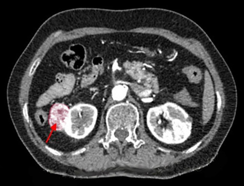
![]() CT Scan (Computed Tomography): The primary imaging method for detecting and staging RCC. Provides detailed images of the kidneys and surrounding tissues.
CT Scan (Computed Tomography): The primary imaging method for detecting and staging RCC. Provides detailed images of the kidneys and surrounding tissues.![]()
![]() MRI (Magnetic Resonance Imaging): May be used to evaluate the tumor further, especially if there are concerns about blood vessel involvement or spread to other organs.
MRI (Magnetic Resonance Imaging): May be used to evaluate the tumor further, especially if there are concerns about blood vessel involvement or spread to other organs.![]()
![]() Ultrasound: Can help differentiate between solid tumors and fluid-filled cysts.
Ultrasound: Can help differentiate between solid tumors and fluid-filled cysts.![]()
![]() Bone Scan: To check for spread to the bones, if suspected.
Bone Scan: To check for spread to the bones, if suspected.![]()
![]() Chest X-ray or CT scan: To look for spread to the lungs.
Chest X-ray or CT scan: To look for spread to the lungs.![]() Biopsy: A sample of kidney tissue is taken and examined under a microscope to confirm the diagnosis and determine the type of RCC. Biopsies are not always necessary, especially if imaging studies are highly suggestive of RCC.
Biopsy: A sample of kidney tissue is taken and examined under a microscope to confirm the diagnosis and determine the type of RCC. Biopsies are not always necessary, especially if imaging studies are highly suggestive of RCC.
Staging:
Staging is crucial for determining the extent of the cancer and guiding treatment decisions. The TNM system is commonly used:![]() T (Tumor): Describes the size and extent of the primary tumor.
T (Tumor): Describes the size and extent of the primary tumor.![]() N (Nodes): Indicates whether the cancer has spread to nearby lymph nodes.
N (Nodes): Indicates whether the cancer has spread to nearby lymph nodes.![]() M (Metastasis): Indicates whether the cancer has spread to distant organs.
M (Metastasis): Indicates whether the cancer has spread to distant organs.
Stages range from I to IV, with Stage I being the least advanced and Stage IV being the most advanced.
Treatment:
Treatment options depend on the stage of the cancer, the patient's overall health, and other factors. Common treatments include:![]() Surgery:
Surgery:![]()
![]() Partial Nephrectomy: Removal of only the tumor and a small margin of healthy kidney tissue. Preferred when possible, especially for smaller tumors and to preserve kidney function.
Partial Nephrectomy: Removal of only the tumor and a small margin of healthy kidney tissue. Preferred when possible, especially for smaller tumors and to preserve kidney function.![]()
![]() Radical Nephrectomy: Removal of the entire kidney, surrounding tissue, and sometimes nearby lymph nodes.
Radical Nephrectomy: Removal of the entire kidney, surrounding tissue, and sometimes nearby lymph nodes.![]() Active Surveillance: For small, slow-growing tumors, especially in elderly or patients with other health problems, monitoring the tumor with regular imaging may be an option instead of immediate treatment.
Active Surveillance: For small, slow-growing tumors, especially in elderly or patients with other health problems, monitoring the tumor with regular imaging may be an option instead of immediate treatment.![]() Ablation Therapies:
Ablation Therapies:![]()
![]() Radiofrequency Ablation (RFA): Uses heat to destroy the tumor.
Radiofrequency Ablation (RFA): Uses heat to destroy the tumor.![]()
![]() Cryoablation: Uses extreme cold to freeze and destroy the tumor.
Cryoablation: Uses extreme cold to freeze and destroy the tumor.![]() Targeted Therapy: Drugs that target specific molecules involved in cancer cell growth and survival. Examples include:
Targeted Therapy: Drugs that target specific molecules involved in cancer cell growth and survival. Examples include:![]()
![]() VEGF inhibitors (e.g., sunitinib, sorafenib, pazopanib, axitinib, cabozantinib): Block the growth of blood vessels that supply the tumor.
VEGF inhibitors (e.g., sunitinib, sorafenib, pazopanib, axitinib, cabozantinib): Block the growth of blood vessels that supply the tumor.![]()
![]() mTOR inhibitors (e.g., temsirolimus, everolimus): Block a protein involved in cell growth and metabolism.
mTOR inhibitors (e.g., temsirolimus, everolimus): Block a protein involved in cell growth and metabolism.![]() Immunotherapy: Drugs that boost the body's immune system to fight the cancer. Examples include:
Immunotherapy: Drugs that boost the body's immune system to fight the cancer. Examples include:![]()
![]() PD-1 inhibitors (e.g., nivolumab, pembrolizumab): Block a protein on immune cells that prevents them from attacking cancer cells.
PD-1 inhibitors (e.g., nivolumab, pembrolizumab): Block a protein on immune cells that prevents them from attacking cancer cells.![]()
![]() CTLA-4 inhibitors (e.g., ipilimumab): Another type of immune checkpoint inhibitor.
CTLA-4 inhibitors (e.g., ipilimumab): Another type of immune checkpoint inhibitor.![]()
![]() IL-2 (Interleukin-2): A cytokine that stimulates the growth and activity of immune cells. Less commonly used due to significant side effects.
IL-2 (Interleukin-2): A cytokine that stimulates the growth and activity of immune cells. Less commonly used due to significant side effects.![]() Radiation Therapy: Uses high-energy rays to kill cancer cells. Less commonly used for RCC because it is often not very effective. May be used to treat bone metastases or to relieve pain.
Radiation Therapy: Uses high-energy rays to kill cancer cells. Less commonly used for RCC because it is often not very effective. May be used to treat bone metastases or to relieve pain.![]() Chemotherapy: Traditional chemotherapy drugs are generally not very effective for RCC.
Chemotherapy: Traditional chemotherapy drugs are generally not very effective for RCC.
Prognosis:
The prognosis for RCC varies depending on the stage of the cancer, the subtype of RCC, and the patient's overall health. Early-stage RCC that is completely removed with surgery has a good prognosis. Advanced-stage RCC has a less favorable prognosis, but newer targeted therapies and immunotherapies have significantly improved outcomes.
Follow-up Care:
After treatment, regular follow-up appointments with your doctor are essential to monitor for recurrence and manage any side effects of treatment. Follow-up may include physical exams, blood tests, and imaging scans.
Important Considerations:![]() Early detection is key: Because early-stage RCC often has no symptoms, regular checkups and awareness of risk factors are important.
Early detection is key: Because early-stage RCC often has no symptoms, regular checkups and awareness of risk factors are important.![]() Personalized treatment: The best treatment approach is tailored to each individual patient based on their specific circumstances.
Personalized treatment: The best treatment approach is tailored to each individual patient based on their specific circumstances.![]() Clinical trials: Participation in clinical trials may offer access to new and promising treatments.
Clinical trials: Participation in clinical trials may offer access to new and promising treatments.
This information is for general knowledge and educational purposes only, and does not constitute medical advice. It is essential to consult with a qualified healthcare professional for any health concerns or before making any decisions related to your health or treatment.