T-cell lymphoma
Published: 18 Jun 2025
ICD9: 202.70 ICD10: C91.50 ICD11: XH3HJ2
T-cell lymphomas (TCLs) are a diverse group of non-Hodgkin lymphomas (NHLs) that develop from abnormal T-lymphocytes (T-cells), a type of white blood cell that plays a crucial role in the immune system.
Unlike B-cell lymphomas, which arise from B-cells, TCLs are less common and often more aggressive.
Here's a breakdown of key aspects of T-cell lymphomas:
What are T-cells?
![]() T-cells are crucial for cell-mediated immunity. They directly attack infected or cancerous cells.
T-cells are crucial for cell-mediated immunity. They directly attack infected or cancerous cells.![]() They mature in the thymus gland.
They mature in the thymus gland.![]() Different types of T-cells include helper T-cells, cytotoxic T-cells, and regulatory T-cells.
Different types of T-cells include helper T-cells, cytotoxic T-cells, and regulatory T-cells.
Key Characteristics of T-Cell Lymphomas:![]() Origin: Develop from mature or immature T-cells that become cancerous.
Origin: Develop from mature or immature T-cells that become cancerous.![]() Rarity: Less common than B-cell lymphomas, accounting for about 15% of all NHLs.
Rarity: Less common than B-cell lymphomas, accounting for about 15% of all NHLs.![]() Heterogeneity: A very diverse group of diseases, meaning there are many different subtypes with varying behaviors and prognoses.
Heterogeneity: A very diverse group of diseases, meaning there are many different subtypes with varying behaviors and prognoses.![]() Aggressiveness: Often more aggressive than B-cell lymphomas. Some types are indolent (slow-growing), but many are aggressive and require immediate treatment.
Aggressiveness: Often more aggressive than B-cell lymphomas. Some types are indolent (slow-growing), but many are aggressive and require immediate treatment.![]() Location: Can affect various parts of the body, including:
Location: Can affect various parts of the body, including:![]()

![]() Lymph nodes: Leading to swollen lymph nodes (lymphadenopathy).
Lymph nodes: Leading to swollen lymph nodes (lymphadenopathy).![]()
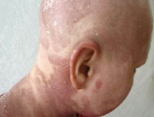
![]() Skin: Leading to skin lesions (cutaneous T-cell lymphoma).
Skin: Leading to skin lesions (cutaneous T-cell lymphoma).![]()
![]() Blood: Leading to leukemia-like symptoms.
Blood: Leading to leukemia-like symptoms.![]()
![]() Bone marrow: Affecting blood cell production.
Bone marrow: Affecting blood cell production.![]()
![]() Other organs: Liver, spleen, gastrointestinal tract, etc.
Other organs: Liver, spleen, gastrointestinal tract, etc.
Types of T-Cell Lymphomas:
There are numerous subtypes of TCLs, and they are generally classified based on:![]() Where they originate (e.g., cutaneous vs. systemic)
Where they originate (e.g., cutaneous vs. systemic)![]() The type of T-cell they derive from (e.g., helper T-cells, cytotoxic T-cells)
The type of T-cell they derive from (e.g., helper T-cells, cytotoxic T-cells)![]() Molecular and genetic features
Molecular and genetic features
Some common subtypes include:![]() Cutaneous T-Cell Lymphomas (CTCL):
Cutaneous T-Cell Lymphomas (CTCL):![]()
![]() Mycosis Fungoides (MF): The most common type of CTCL; primarily affects the skin.
Mycosis Fungoides (MF): The most common type of CTCL; primarily affects the skin.![]()
![]() Sézary Syndrome (SS): A leukemic form of CTCL, characterized by widespread skin involvement, enlarged lymph nodes, and abnormal T-cells (Sézary cells) in the blood.
Sézary Syndrome (SS): A leukemic form of CTCL, characterized by widespread skin involvement, enlarged lymph nodes, and abnormal T-cells (Sézary cells) in the blood.![]() Peripheral T-Cell Lymphomas (PTCL): These arise from mature T-cells.
Peripheral T-Cell Lymphomas (PTCL): These arise from mature T-cells.![]()
![]() Anaplastic Large Cell Lymphoma (ALCL): Some types are associated with a protein called ALK (anaplastic lymphoma kinase).
Anaplastic Large Cell Lymphoma (ALCL): Some types are associated with a protein called ALK (anaplastic lymphoma kinase).![]()
![]() Angioimmunoblastic T-Cell Lymphoma (AITL): Often associated with immune system abnormalities.
Angioimmunoblastic T-Cell Lymphoma (AITL): Often associated with immune system abnormalities.![]()
![]() Peripheral T-Cell Lymphoma, Not Otherwise Specified (PTCL-NOS): A diagnosis used when the lymphoma doesn't fit into a specific category.
Peripheral T-Cell Lymphoma, Not Otherwise Specified (PTCL-NOS): A diagnosis used when the lymphoma doesn't fit into a specific category.![]()
![]() Adult T-Cell Leukemia/Lymphoma (ATLL): Caused by the Human T-lymphotropic virus type 1 (HTLV-1). More common in certain geographic regions.
Adult T-Cell Leukemia/Lymphoma (ATLL): Caused by the Human T-lymphotropic virus type 1 (HTLV-1). More common in certain geographic regions.![]()
![]() Extranodal NK/T-Cell Lymphoma, Nasal Type: Often associated with Epstein-Barr virus (EBV) and commonly found in the nasal cavity and upper respiratory tract.
Extranodal NK/T-Cell Lymphoma, Nasal Type: Often associated with Epstein-Barr virus (EBV) and commonly found in the nasal cavity and upper respiratory tract.![]() Precursor T-Lymphoblastic Lymphoma/Leukemia: This is more commonly diagnosed as acute lymphoblastic leukemia and is seen more in children and young adults.
Precursor T-Lymphoblastic Lymphoma/Leukemia: This is more commonly diagnosed as acute lymphoblastic leukemia and is seen more in children and young adults.
Symptoms:
Symptoms vary depending on the type and location of the lymphoma. Common symptoms include:![]() Swollen lymph nodes: Usually painless.
Swollen lymph nodes: Usually painless.![]() Skin rashes or lesions: Itching, red, scaly patches, or tumors.
Skin rashes or lesions: Itching, red, scaly patches, or tumors.![]() Fatigue
Fatigue![]() Unexplained weight loss
Unexplained weight loss![]() Fever
Fever![]() Night sweats
Night sweats![]() Enlarged liver or spleen
Enlarged liver or spleen![]() Cough or shortness of breath (if affecting the chest)
Cough or shortness of breath (if affecting the chest)
Causes and Risk Factors:
The exact causes of most T-cell lymphomas are unknown. However, some risk factors include:![]() Viral infections: HTLV-1 (Adult T-cell leukemia/lymphoma) and EBV (Extranodal NK/T-Cell Lymphoma, Nasal Type).
Viral infections: HTLV-1 (Adult T-cell leukemia/lymphoma) and EBV (Extranodal NK/T-Cell Lymphoma, Nasal Type).![]() Compromised immune system: Autoimmune disorders or immunosuppressant medications.
Compromised immune system: Autoimmune disorders or immunosuppressant medications.![]() Genetic factors: Some genetic mutations may increase the risk.
Genetic factors: Some genetic mutations may increase the risk.![]() Age: Some subtypes are more common in older adults.
Age: Some subtypes are more common in older adults.![]() Exposure to certain chemicals: Possibly, but the link is not always clear.
Exposure to certain chemicals: Possibly, but the link is not always clear.
Diagnosis:
Diagnosis typically involves:![]() Physical exam: To check for swollen lymph nodes and other signs of the disease.
Physical exam: To check for swollen lymph nodes and other signs of the disease.![]() Lymph node biopsy: Removing a lymph node for microscopic examination.
Lymph node biopsy: Removing a lymph node for microscopic examination.![]() Skin biopsy: If skin lesions are present.
Skin biopsy: If skin lesions are present.![]() Blood tests: Complete blood count (CBC), blood chemistry, and tests for viral infections.
Blood tests: Complete blood count (CBC), blood chemistry, and tests for viral infections.![]() Bone marrow biopsy: To assess bone marrow involvement.
Bone marrow biopsy: To assess bone marrow involvement.![]() Imaging tests: CT scans, PET scans, and MRI scans to determine the extent of the disease.
Imaging tests: CT scans, PET scans, and MRI scans to determine the extent of the disease.![]() Flow cytometry: A technique used to identify and count specific types of cells in the blood or bone marrow.
Flow cytometry: A technique used to identify and count specific types of cells in the blood or bone marrow.![]() Molecular testing: To identify specific genetic mutations or markers that may help with diagnosis and treatment.
Molecular testing: To identify specific genetic mutations or markers that may help with diagnosis and treatment.
Treatment:
Treatment depends on the specific type and stage of the lymphoma, as well as the patient's overall health. Common treatment options include:![]() Chemotherapy: Using drugs to kill cancer cells.
Chemotherapy: Using drugs to kill cancer cells.![]() Radiation therapy: Using high-energy rays to kill cancer cells.
Radiation therapy: Using high-energy rays to kill cancer cells.![]() Stem cell transplant: Replacing damaged bone marrow with healthy stem cells (either from the patient or a donor).
Stem cell transplant: Replacing damaged bone marrow with healthy stem cells (either from the patient or a donor).![]() Targeted therapy: Using drugs that target specific molecules involved in cancer cell growth and survival. Examples include:
Targeted therapy: Using drugs that target specific molecules involved in cancer cell growth and survival. Examples include:![]()
![]() Brentuximab vedotin: An antibody-drug conjugate used for ALCL and other CD30-positive TCLs.
Brentuximab vedotin: An antibody-drug conjugate used for ALCL and other CD30-positive TCLs.![]()
![]() Romidepsin and Vorinostat: Histone deacetylase (HDAC) inhibitors used for CTCL and PTCL.
Romidepsin and Vorinostat: Histone deacetylase (HDAC) inhibitors used for CTCL and PTCL.![]()
![]() Pralatrexate: A folate analog used for PTCL.
Pralatrexate: A folate analog used for PTCL.![]() Immunotherapy: Using drugs to boost the body's immune system to fight cancer cells.
Immunotherapy: Using drugs to boost the body's immune system to fight cancer cells.![]() Photopheresis: A treatment for CTCL (especially Sézary syndrome) where blood is treated with ultraviolet light.
Photopheresis: A treatment for CTCL (especially Sézary syndrome) where blood is treated with ultraviolet light.![]() Skin-directed therapies: For CTCL, these can include topical steroids, retinoids, and other medications applied directly to the skin.
Skin-directed therapies: For CTCL, these can include topical steroids, retinoids, and other medications applied directly to the skin.
Prognosis:
The prognosis for T-cell lymphomas varies widely depending on the specific subtype, stage, and the patient's overall health. Some types are more aggressive and have a poorer prognosis than others. Early diagnosis and treatment are crucial for improving outcomes. Clinical trials are ongoing to develop new and more effective treatments for T-cell lymphomas.
In Summary:
T-cell lymphomas are a diverse and often challenging group of cancers. Understanding the specific subtype of lymphoma, the stage of the disease, and the available treatment options is crucial for effective management and improving the patient's prognosis. It's vital to consult with a hematologist or oncologist experienced in treating lymphomas for accurate diagnosis and personalized treatment planning.